Share to: share facebook share twitter share wa share telegram print page

Midbrain

Midbrain
Figure shows the midbrain (A) and surrounding regions; sagittal view of one cerebellar hemisphere. B: Pons. C: Medulla. D: Spinal cord. E: Fourth ventricle. F: Arbor vitae. G: Nodule. H: Tonsil. I: Posterior lobe. J: Anterior lobe. K: Inferior colliculus. L: Superior colliculus.
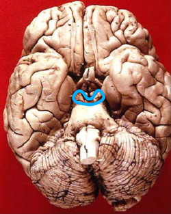
Inferior view in which the midbrain is encircled blue.
Details
PronunciationUK: /ˌmɛsɛnˈsɛfəlɒn, -kɛf-/, US: /ˌmɛzənˈsɛfələn/;[1]
Part ofBrainstem
Identifiers
Latinmesencephalon
MeSHD008636
NeuroNames462
NeuroLex IDbirnlex_1667
TA98A14.1.03.005
TA25874
FMA61993
Anatomical terms of neuroanatomy

The midbrain or mesencephalon is the uppermost portion of the brainstem connecting the diencephalon and cerebrum with the pons.[2] It consists of the cerebral peduncles, tegmentum, and tectum.

It is functionally associated with vision, hearing, motor control, sleep and wakefulness, arousal (alertness), and temperature regulation.[3]

The name mesencephalon comes from the Greek mesos, "middle", and enkephalos, "brain".[4]

Structure

Brainstem (dorsal view)
A:Thalamus B:Midbrain C:Pons
D:Medulla oblongata
7 and 8 are the four colliculi.

The midbrain is the shortest segment of the brainstem, measuring less than 2cm in length. It is situated mostly in the posterior cranial fossa, with its superior part extending above the tentorial notch.[2]

The principal regions of the midbrain are the tectum, the cerebral aqueduct, tegmentum, and the cerebral peduncles. Rostrally the midbrain adjoins the diencephalon (thalamus, hypothalamus, etc.), while caudally it adjoins the hindbrain (pons, medulla and cerebellum).[5] In the rostral direction, the midbrain noticeably splays laterally.

The midbrain is typically sectioned axially at either the superior or inferior colliculi levels. Visualizing these cross-sections as an upside-down bear face helps remember its structures, with the peduncles forming ears, aqueducts mouth, and tectum chin.

Tectum

Principal connections of the tectum

The tectum (Latin for roof) is the part of the midbrain dorsal to the cerebral aqueduct.[2] The position of the tectum is contrasted with the tegmentum, which refers to the region in front of the ventricular system, or floor of the midbrain.

It is involved in certain reflexes in response to visual or auditory stimuli. The reticulospinal tract, which exerts some control over alertness, takes input from the tectum,[6] and travels both rostrally and caudally from it.

The corpora quadrigemina are four mounds, called colliculi, in two pairs – a superior and an inferior pair, on the surface of the tectum. The superior colliculi process some visual information, aid the decussation of several fibres of the optic nerve (some fibres remain ipsilateral), and are involved with saccadic eye movements. The tectospinal tract connects the superior colliculi to the cervical nerves of the neck, and co-ordinates head and eye movements. Each superior colliculus also sends information to the corresponding lateral geniculate nucleus, with which it is directly connected. The homologous structure to the superior colliculus in non mammalian vertebrates including fish and amphibians, is called the optic tectum; in those animals, the optic tectum integrates sensory information from the eyes and certain auditory reflexes.[7][8]

The inferior colliculi – located just above the trochlear nerve – process certain auditory information. Each inferior colliculus sends information to the corresponding medial geniculate nucleus, with which it is directly connected.

Cerebral aqueduct

Ventricular system anatomy showing the cerebral aqueduct, labelled centre right.

The cerebral aqueduct is the part of the ventricular system which links the third ventricle (rostrally) with the fourth ventricle (caudally); as such it is responsible for continuing the circulation of cerebrospinal fluid. The cerebral aqueduct is a narrow channel located between the tectum and the tegmentum, and is surrounded by the periaqueductal grey,[9] which has a role in analgesia, quiescence, and bonding. The dorsal raphe nucleus (which releases serotonin in response to certain neural activity) is located at the ventral side of the periaqueductal grey, at the level of the inferior colliculus.

The nuclei of two pairs of cranial nerves are similarly located at the ventral side of the periaqueductal grey – the pair of oculomotor nuclei (which control the eyelid, and most eye movements) is located at the level of the superior colliculus,[10] while the pair of trochlear nuclei (which helps focus vision on more proximal objects) is located caudally to that, at the level of the inferior colliculus, immediately lateral to the dorsal raphe nucleus.[9] The oculomotor nerve emerges from the nucleus by traversing the ventral width of the tegmentum, while the trochlear nerve emerges via the tectum, just below the inferior colliculus itself; the trochlear is the only cranial nerve to exit the brainstem dorsally. The Edinger-Westphal nucleus (which controls the shape of the lens and size of the pupil) is located between the oculomotor nucleus and the cerebral aqueduct.[9]

Tegmentum

Cross-section of the midbrain at the level of the superior colliculus
Cross-section of the midbrain at the level of the inferior colliculus.

The midbrain tegmentum is the portion of the midbrain ventral to the cerebral aqueduct, and is much larger in size than the tectum. It communicates with the cerebellum by the superior cerebellar peduncles, which enter at the caudal end, medially, on the ventral side; the cerebellar peduncles are distinctive at the level of the inferior colliculus, where they decussate, but they dissipate more rostrally.[9] Between these peduncles, on the ventral side, is the median raphe nucleus, which is involved in memory consolidation.

The main bulk of the tegmentum contains a complex synaptic network of neurons, primarily involved in homeostasis and reflex actions. It includes portions of the reticular formation. A number of distinct nerve tracts between other parts of the brain pass through it. The medial lemniscus – a narrow ribbon of fibres – passes through in a relatively constant axial position; at the level of the inferior colliculus it is near the lateral edge, on the ventral side, and retains a similar position rostrally (due to widening of the tegmentum towards the rostral end, the position can appears more medial). The spinothalamic tract – another ribbon-like region of fibres – are located at the lateral edge of the tegmentum; at the level of the inferior colliculus it is immediately dorsal to the medial lemiscus, but due to the rostral widening of the tegmentum, is lateral of the medial lemiscus at the level of the superior colliculus.

A prominent pair of round, reddish, regions – the red nuclei (which have a role in motor co-ordination) – are located in the rostral portion of the midbrain, somewhat medially, at the level of the superior colliculus.[9] The rubrospinal tract emerges from the red nucleus and descends caudally, primarily heading to the cervical portion of the spine, to implement the red nuclei's decisions. The area between the red nuclei, on the ventral side – known as the ventral tegmental area – is the largest dopamine-producing area in the brain, and is heavily involved in the neural reward system. The ventral tegmental area is in contact with parts of the forebrain – the mammillary bodies (from the Diencephalon) and hypothalamus (of the diencephalon).

Cerebral peduncles

Brain anatomy – forebrain, midbrain, hindbrain.

The cerebral peduncles each form a lobe ventrally of the tegmentum, on either side of the midline. Beyond the midbrain, between the lobes, is the interpeduncular fossa, which is a cistern filled with cerebrospinal fluid [citation needed].

The majority of each lobe constitutes the cerebral crus. The cerebral crus are the main tracts descending from the thalamus to caudal parts of the central nervous system; the central and medial ventral portions contain the corticobulbar and corticospinal tracts, while the remainder of each crus primarily contains tracts connecting the cortex to the pons. Older texts refer to the crus cerebri as the cerebral peduncle; however, the latter term actually covers all fibres communicating with the cerebrum (usually via the diencephalon), and therefore would include much of the tegmentum as well. The remainder of the crus pedunculi – small regions around the main cortical tracts – contain tracts from the internal capsule.

The portion of the lobes in connection with the tegmentum, except the most lateral portion, is dominated by a blackened band – the substantia nigra (literally black substance)[9] – which is the only part of the basal ganglia system outside the forebrain. It is ventrally wider at the rostral end. By means of the basal ganglia, the substantia nigra is involved in motor-planning, learning, addiction, and other functions. There are two regions within the substantia nigra – one where neurons are densely packed (the pars compacta) and one where they are not (the pars reticulata), which serve a different role from one another within the basal ganglia system. The substantia nigra has extremely high production of melanin (hence the colour), dopamine, and noradrenalin; the loss of dopamine-producing neurons in this region contributes to the progression of Parkinson's disease.[11]

Blood supply

The midbrain is supplied by the following arteries:

Venous blood from the midbrain is mostly drained into the basal vein as it passes around the peduncle. Some venous blood from the colliculi drains to the great cerebral vein.[12]

Development

Mesencephalon of human embryo

During embryonic development, the midbrain (also known as the mesencephalon) arises from the second vesicle of the neural tube, while the interior of this portion of the tube becomes the cerebral aqueduct. Unlike the other two vesicles – the forebrain and hindbrain – the midbrain does not develop further subdivision for the remainder of neural development. It does not split into other brain areas. While the forebrain, for example, divides into the telencephalon and the diencephalon.[13]

Throughout embryonic development, the cells within the midbrain continually multiply; this happens to a much greater extent ventrally than it does dorsally. The outward expansion compresses the still-forming cerebral aqueduct, which can result in partial or total obstruction, leading to congenital hydrocephalus.[14] The tectum is derived in embryonic development from the alar plate of the neural tube.

Function

The midbrain is the uppermost part of the brainstem. Its substantia nigra is closely associated with motor system pathways of the basal ganglia. The human midbrain is archipallian in origin, meaning that its general architecture is shared with the most ancient of vertebrates. Dopamine produced in the substantia nigra and ventral tegmental area plays a role in movement, movement planning, excitation, motivation and habituation of species from humans to the most elementary animals such as insects. Laboratory mice from lines that have been selectively bred for high voluntary wheel running have enlarged midbrains.[15] The midbrain helps to relay information for vision and hearing.

The term "tectal plate" or "quadrigeminal plate" is used to describe the junction of the gray and white matter in the embryo. (ancil-453 at NeuroNames)

See also

References

  1. ^ "mesencephalon". Oxford English Dictionary (Online ed.). Oxford University Press. (Subscription or participating institution membership required.)
  2. ^ a b c Sinnatamby, Chummy S. (2011). Last's Anatomy (12th ed.). p. 476. ISBN 978-0-7295-3752-0.
  3. ^ Breedlove, Watson, & Rosenzweig. Biological Psychology, 6th Edition, 2010, pp. 45-46
  4. ^ Mosby's Medical, Nursing & Allied Health Dictionary,≈ Fourth Edition, Mosby-Year Book 1994, p. 981
  5. ^ "Slide 5". Archived from the original on 2011-04-27. Retrieved 2011-03-05.
  6. ^ Kandel, Eric (2000). Principles of Neural Science. McGraw-Hill. pp. 669. ISBN 0-8385-7701-6.
  7. ^ Collins Dictionary of Biology, 3rd ed. © W. G. Hale, V. A. Saunders, J. P. Margham 2005
  8. ^ Ferrier, David (1886). "Functions of the optic lobes or corpora quadrigemina": 149–173. doi:10.1037/12789-005. {{cite journal}}: Cite journal requires |journal= (help)
  9. ^ a b c d e f Martin. Neuroanatomy Text and Atlas, Second edition. 1996, pp. 522-525.
  10. ^ Haines, Duane E. (2012). Neuroanatomy : an atlas of structures, sections, and systems (8th ed.). Philadelphia: Wolters Kluwer/ Lippincott Williams & Wilkins Health. pp. 42. ISBN 978-1-60547-653-7.
  11. ^ Damier, P.; Hirsch, E. C.; Agid, Y.; Graybiel, A. M. (1999-08-01). "The substantia nigra of the human brainII. Patterns of loss of dopamine-containing neurons in Parkinson's disease". Brain. 122 (8): 1437–1448. doi:10.1093/brain/122.8.1437. ISSN 0006-8950. PMID 10430830.
  12. ^ Sinnatamby, Chummy S. (2011). Last's Anatomy (12th ed.). p. 478. ISBN 978-0-7295-3752-0.
  13. ^ Martin. Neuroanatomy Text and Atlas, Second Edition, 1996, pp. 35-36.
  14. ^ "Hydrocephalus Fact Sheet". National Institute of Neurological Disorders and Stroke. February 2008. Retrieved 2011-03-23.
  15. ^ Kolb, E. M.; Rezende, E. L.; Holness, L.; Radtke, A.; Lee, S. K.; Obenaus, A.; Garland (2013). "Mice selectively bred for high voluntary wheel running have larger midbrains: support for the mosaic model of brain evolution". Journal of Experimental Biology. 216 (3): 515–523. doi:10.1242/jeb.076000. PMID 23325861.

Read other articles:

هذه المقالة يتيمة إذ تصل إليها مقالات أخرى قليلة جدًا. فضلًا، ساعد بإضافة وصلة إليها في مقالات متعلقة بها. (ديسمبر 2021) حرب السهام최종병기 활 (بالكورية) معلومات عامةالصنف الفني فيلم أكشن[1][2] — فيلم حربي — فيلم دراما تاريخ الصدور 10 أغسطس 2011[3] مدة العرض 122 دقيقة اللغة ا


2016 book by Claudia Casper The Mercy Journals First edition coverAuthorClaudia CasperCountryCanadaLanguageEnglishGenrePost-apocalyptic fiction, science fictionPublisherArsenal Pulp PressPublication date2016Media typePrint, ebookPages234ISBN9781551526331Dewey Decimal813.6LC ClassPR9199.3.C4315 M47 2016 The Mercy Journals is a 2016 post-apocalyptic science fiction novel by Canadian author Claudia Casper. The novel, set in a near-future world where the global population has been reduc...


Медаль «Золота Зірка» Список Національних Героїв Азербайджану Список Це неповний список відомих Національних героїв: Дата Фото ПрізвищеІм'я Друге ім'я Дата народження Місце народження Дата смерті Місце смерті 7 липня 1992 Агарунов Альберт Агарунович 25 квітня 1969 пос....

Oum er-Rbia Quellgebiet des Oum er-Rbia Quellgebiet des Oum er-Rbia Daten Lage Marokko Marokko Flusssystem Oum er-Rbia Quelle Mittlerer Atlas, ca. 40 km nordöstlich von Khenifra33° 1′ 1″ N, 5° 19′ 12″ W33.017-5.32011613 Quellhöhe 1613 m Mündung nördlich von Azemmour in den Atlantik33.3191-8.33780Koordinaten: 33° 19′ 9″ N, 8° 20′ 16″ W33° 19′ 9″ N...


Cricket tournament 2021–22 Major Clubs Limited Over TournamentDates27 October – 30 November 2021Administrator(s)Sri Lanka CricketCricket formatList A cricketTournament format(s)Round-robin then knockoutHost(s) Sri LankaChampionsTamil Union Cricket and Athletic ClubParticipants26Most runsChanaka Ruwansiri (320)Most wicketsJeffrey Vandersay (24)← 2020–21 The 2021–22 Major Clubs Limited Over Tournament was a List A cricket competition that took place in Sri Lanka.[1] I...


Bagian dari seriGereja Katolik menurut negara Afrika Afrika Selatan Afrika Tengah Aljazair Angola Benin Botswana Burkina Faso Burundi Chad Eritrea Eswatini Etiopia Gabon Gambia Ghana Guinea Guinea-Bissau Guinea Khatulistiwa Jibuti Kamerun Kenya Komoro Lesotho Liberia Libya Madagaskar Malawi Mali Maroko Mauritania Mauritius Mesir Mozambik Namibia Niger Nigeria Pantai Gading Republik Demokratik Kongo Republik Kongo Rwanda Sao Tome dan Principe Senegal Seychelles Sierra Leone Somalia Somaliland ...

Premier of Saskatchewan from 1926 to 1929 and 1934 to 1935 The Right HonourableJames Garfield GardinerPC4th Premier of SaskatchewanIn officeFebruary 26, 1926 – September 9, 1929MonarchGeorge VLieutenant GovernorHenry William NewlandsPreceded byCharles A. DunningSucceeded byJames T.M. AndersonIn officeJuly 19, 1934 – November 1, 1935MonarchGeorge VLieutenant GovernorHugh Edwin MunroePreceded byJames T.M. AndersonSucceeded byWilliam John PattersonMember of the Legislat...


Chùa Phật Lớn(Thiên Trúc tự)Vị tríQuốc gia Việt NamĐịa chỉ197/11 đường Phương Thành, phường Bình San, thành phố Hà Tiên, tỉnh Kiên Giang, Việt NamThông tinTôn giáoPhật giáoKhởi lậptrước thế kỷ 16 Cổng thông tin Phật giáoxts Đối với chùa cùng tên tại Việt Nam, xem chùa Thiên Trúc. Đối với chùa cùng tên tại Việt Nam, xem chùa Phật Lớn. Bài viết này cần thêm chú thích nguồn gốc đ


フィリップス男爵ニコラス・フィリップス(2018年3月) ワース・マトラヴァーズのフィリップス男爵、ニコラス・アディソン・フィリップス(英: Nicholas Addison Phillips, Baron Phillips of Worth Matravers, KG, PC、1938年1月2日 -)は、イギリスの裁判官、法服貴族、政治家。 裁判官としてキャリアを積み、1999年に常任上訴法服貴族(英語版)となり、2008年から首席常任上訴法服貴族

Kedutaan Besar Republik Indonesia di VientianeKoordinat17°58′24″N 102°37′20″E / 17.973323°N 102.622331°E / 17.973323; 102.622331Lokasi Vientiane, LaosAlamatKaysone Phomvihane Ave.Vientiane, LaosDuta BesarGrata Endah WerdaningtyasYurisdiksi LaosSitus webkemlu.go.id/vientiane/id Kedutaan Besar Republik Indonesia di Vientiane (KBRI Vientiane) adalah misi diplomatik Republik Indonesia untuk Republik Demokratik Rakyat Laos.[1] Daftar duta besar Arti...


Railway station in Damansara Town Centre, Malaysia  KG13  Pusat Bandar Damansara Pavilion Damansara Heights – Pusat Bandar Damansara | MRT stationPusat Bandar Damansara MRT stationGeneral informationOther namesChinese: 白沙罗市中心Tamil: சென்டர் பெருநகரம் டாமன்சாராLocationDamansara Town Centre, Damansara,Kuala Lumpur, Malaysia.Coordinates3°8′36.28″N 101°39′44.07″E / 3.1434111°N 101.6622417°E&#x...


2006 video gameScooby-Doo! Who's Watching Who?North American PSP cover artDeveloper(s)Savage Entertainment (PSP)Human Soft, Inc. (DS)Publisher(s)THQPlatform(s)PlayStation Portable, Nintendo DSReleaseNA: October 11, 2006[1][2]EU: November 17, 2006AU: November 23, 2006Genre(s)Action-adventureMode(s)Single-player Scooby-Doo! Who's Watching Who? is a third-person action-adventure video game developed by Savage Entertainment and Human Soft, Inc. and published by THQ for the PlaySta...

Margaret DilkeBornMargaret Mary Smith4 September 1857Died19 May 1914 (1914-05-20) (aged 56)Newport, Isle of Wight, England, United Kingdom of Great Britain and IrelandNationalityBritishOther namesMrs. William Russell CookeOccupationcampaigner for women's rightsSpouses Ashton Wentworth Dilke ​ ​(m. 1876; died 1883)​ William Russell Cooke ​ ​(m. 1891)​ Margaret Maye Dilke born Margaret Mary Smith b...


36th President of Guatemala from 1974 to 1978 In this Spanish name, the first or paternal surname is Laugerud and the second or maternal family name is García. GeneralKjell Eugenio Laugerud GarcíaOfficial portrait, 197436th President of GuatemalaIn office1 July 1974 (1974-07-01) – 1 July 1978 (1978-07-01)Vice PresidentMario Sandoval AlarcónPreceded byCarlos Arana OsorioSucceeded byFernando Romeo Lucas García Personal detailsBorn(1930-01-24)...


Lo Morant-San Nicolás de Bari Barrio El parque Lo Morant Coordenadas 38°22′04″N 0°29′25″O / 38.367861111111, -0.49025Entidad Barrio • País  España • Ciudad Alicante • Provincia Alicante • CC.AA. Comunidad ValencianaPoblación (2022)   • Total 6755 hab.Código postal 03010[editar datos en Wikidata] Lo Morant-San Nicolás de Bari es un barrio de la ciudad española de Alicante. Según el padrón munici...

Este artigo ou secção contém uma lista de referências no fim do texto, mas as suas fontes não são claras porque não são citadas no corpo do artigo, o que compromete a confiabilidade das informações. Ajude a melhorar este artigo inserindo citações no corpo do artigo. (Junho de 2021) Fajã das Almas, Ermida de Santo Cristo, Manadas. Fajã das Almas, Igreja de Santo Cristo, interior. A Ermida de Santo Cristo é uma ermida Portuguesa localizada na Fajã das Almas, a freguesia da Manad...


Species of bird Russet-crowned quail-dove Conservation status Near Threatened (IUCN 3.1)[1] Scientific classification Domain: Eukaryota Kingdom: Animalia Phylum: Chordata Class: Aves Order: Columbiformes Family: Columbidae Genus: Zentrygon Species: Z. goldmani Binomial name Zentrygon goldmani(Nelson, 1912) Synonyms Geotrygon goldmani Oreopelia goldmani The russet-crowned quail-dove (Zentrygon goldmani) is a species of bird in the family Columbidae. It is found in Panama and ...


This article uses bare URLs, which are uninformative and vulnerable to link rot. Please consider converting them to full citations to ensure the article remains verifiable and maintains a consistent citation style. Several templates and tools are available to assist in formatting, such as reFill (documentation) and Citation bot (documentation). (September 2022) (Learn how and when to remove this template message) Ernest Hemingway with his family and four marlin in 1935 Marlin fishing or billf...

Sugarland Mountain TrailSugarland Mountain Trailhead at Fighting Creek GapLength12 mi (19 km)LocationGreat Smoky Mountains National Park, Tennessee, United StatesTrailheadsFighting Creek Gap (along Little River Road, W. of Gatlinburg)Junction with the Appalachian Trail near the summit of Mount CollinsUseHikingHighest pointAppalachian Trail junction, 5,900 ft (1,800 m)Lowest pointFighting Creek Gap, 2,300 ft (700 m)DifficultyModerate-to-StrenuousSeasonOpen year-ro...


Kucing berkaki pendek ras Munchkin. Kucing kerdil atau kucing katai adalah kucing domestik yang memiliki kondisi dwarfisme (kekerdilan) yang disebabkan oleh mutasi genetik. Tidak seperti kucing dengan proporsi normal, kucing kerdil akan memperlihatkan gejala osteokondrodisplasia, yaitu kelainan genetik pada tulang dan tulang rawan, sehingga kaki menjadi terlihat pendek.[1] Sejak pertengahan abad ke-20, ras kucing dengan kelainan dwarfisme telah dikembangkan untuk penjualan komersial. ...

 
Kembali kehalaman sebelumnya